Tuesday, November 29, 2011

Whiplash - Cervical Zygapophysial Joint Pain After Whiplash

From: On Cervical Zygapophysial Joint Pain After Whiplash. Bogduk, Nikolai MD, PhD. Spine: 1 December 2011 - Volume 36 - Issue - p S194–S199.

What is Whiplash ? 

Whiplash is an injury to the neck and upper back that can occur following an auto accident. Whiplash can also occur in other circumstances where the neck is forced backwards and then forwards such as a fall or during a sporting event. In automobile accidents, whiplash occurs as a result of sudden "acceleration - deceleration" forced to the spine and soft tissue structures (tendons, ligaments and muscles):

Whiplash Associated Disorder (WAD), describes a more serious and chronic condition which can include symptoms other than just pain and stiffness in the neck such as: nausea, dizziness, headaches, ringing in the ears, concussion, etc. 

In automobile accidents, the most common form of impact for whiplash is a rear end type of impact. There are many factors contributing to the injury including: speed of impact, type of seat, type of vehicle, did the occupant brace for impact ? was the occupant looking straight ahead or turned ? etc.

Whiplash can be quantified on X-ray as an "S-Shaped curve": 

It has been well documented that soft tissue structures are injured and which are responsible for localized pain and stiffness. Pain and tingling down the arm(s) can be a sign of a more serious condition such as a cervical disc herniation or a brachial plexus lesion or compression. 

This recent study suggests what Chiropractors have always known, that there is injury to cervical zygapophysial joints OR facet joints following whiplash. Facet joints are located at each level of our vertebrae and are the joints responsible for the "popping or crackling sounds" that we hear from our spines. Facet joints are also the joints that are "adjusted" when Chiropractors manipulate the spine. Perhaps that is why so many patients have found relief with Chiropractic following an automobile accident causing whiplash

Diagram showing Facet joints:

Have you been injured in an automobile accident and are suffering from whiplash, headaches or an injury to a spinal disc and nerve ? Call us at (650) 347-2225 to get a correct diagnosis and appropriate treatment. We offer treatment for soft tissue injuries, injuries to the facet joints and cervical disc's. Dr. Hindocha is a Chiropractor in San Mateo who has had over 10 years of experience managing these difficult cases. Moreover, Dr. Hindocha shares his office space with a Physical Medicine & Rehabilitation and Orthopedic specialist should you require advanced care. We offer: Chiropractic care, non-surgical spinal decompression therapy, PT modalities and procedures, therapeutic exercises such as cervical and lumbar core stabilization programs and traction procedures. We will work with your lawyer or can recommend an attorney qualified for your case. Dr. Hindocha's paperwork and medical reports can help support your case to avoid costly litigation.

Assessing the outcomes after Spinal Fusion surgery.

From: Perioperative Morbidity and Mortality After Anterior, Posterior, and Anterior/Posterior Spine Fusion Surgery. Stavros G. Memtsoudis, MD, PhD; Vassilios I. Vougioukas, MD, PhD; Yan Ma, PhD; Licia K. Gaber-Baylis, BA; Federico P. Girardi, MD. Spine. 2011;36(22):1867-1877. © 2011 Lippincott Williams & Wilkins.

The utilization of Spinal fusion techniques have increased dramatically despite the relatively few studies showing the safety of this procedure. There is also a relative lack of studies comparing the relative spinal surgical techniques and the outcomes associated with them.


This study was designed to assess the Morbidity (the rate of incidence) and Mortality (the measure of the number of deaths) and to determine the independent risk factors for in-hospital death. associated with the various surgical approaches to spinal fusion surgery.

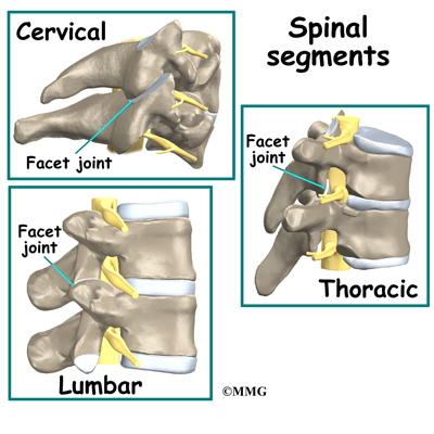
The outcomes being measured were: Perioperative (after operation) of Anterior Spinal Fusion (ASF), Posterior Spinal Fusion (PSF) and APSF (Anterior Posterior Spinal Fusion). 


Here are some X-rays, post-operative:


Results of the study:


There was an increased incidence of perioperative complications and adjusted risk of in-hospital mortality among hospital admissions undergoing APSF and ASF when compared to PSF procedures.

The highest rates of fatal outcomes and complications were associated with procedures using the anterior thoracic approach.

Risk factors for in-hospital mortality included the following: male gender, advanced age, procedures indicated for metastatic disease and trauma, as well as the presence of several comorbidities and perioperative complications.

Procedures involving the anterior spine were associated with higher morbidity and mortality in our study, despite being performed in younger individuals with lower comorbidity burden.

The highest rate of morbidity and mortality was seen in APSF patients, which can be explained by longer surgical times, more blood loss, and increased surgical complexity. 

When studying patient demographics and their association with mortality, we found increased independent risk of a fatal event after spine fusion among men.

We identified an increased incidence of morbidity and risk for mortality in patients with advanced age. Patients over the age of 75 years made up almost one- third of all mortalities, despite representing less than 9% of the spine surgical population in this study.

Pulmonary circulatory disease, congestive heart failure, renal disease, and coagulopathies were associated with the highest increases of risk for perioperative mortality.

Perioperative complications were also associated with increases in the odds of a fatal event. Pulmonary embolism, perioperative shock, ARDS, and cardiac complications were associated with the highest risk of mortality. All of these events had the highest incidence among APSF patients.

It was determined that APSF and ASF carried an increased adjusted risk of in-hospital mortality and greater incidence of in-hospital complications when compared to PSF procedures.

What can we learn from this study ? Like the old saying goes "an ounce of prevention equals a pound of cure". Aside from unforeseen circumstances such as trauma, there are ways we can help prevent our spines from getting to this level of degeneration.

1.) Have a spinal exam and check up.

2.) Don't ignore early signs such as: recurring back pain that may self-resolve or resolve with medications. In my experience, most cases of spinal degeneration and herniated discs have manifested themselves in the past and have not been adequately or properly treated. Chiropractic therapy is a drugless, non-surgical, intervention which can help with many types of back pain.

3.) For those of you who have already reached the advanced stages of spinal arthritis, osteroarthritis, degenerative disc disease, sciatica or have one or multiple herniated discs, VAX-D spinal decompression therapy is an alternative approach to some types of surgery and "endless" epidural injections.

4.) Exercise, lose weight and keep a healthy lifestyle.

Dr. Rommel Hindocha is a Chiropractor in San Mateo California. In addition to Chiropractic therapy, he does perform non-surgical Spinal Decompression therapy at 101 S. San Mateo Drive, Suite 200, San Mateo, CA  94401. You can reach Peninsula Spine & Sports Rehabilitation at (650) 347-2225.


Wednesday, November 2, 2011

Is there a genetic predisposition to "Back Disease" ?

J Bone Joint Surg Am. 2011;93:225-229. Abstract

In an analysis of a database of more than 2 million people, first-degree and third-degree relatives of people with lumbar disc disease had a significantly increased relative risk of developing the back condition themselves compared with expected rates for the general population. "The results of this study support a heritable predisposition to lumbar disc disease," lead author Alpesh A. Patel, MD, and colleagues from the departments of Orthopaedics and Biomedical Informatics, University of Utah School of Medicine, Salt Lake City, report in the February 2 issue of the Journal of Bone and Joint Surgery.

To test the hypothesis that lumbar disc disease may be inherited, the authors analyzed data from both the Utah Population Database, which permits the tracking of medical information on the founding pioneers of Utah and their descendents, and the University of Utah Health Sciences Center data warehouse, which has diagnosis and procedure data on all patients treated at the University Hospital. Together, the databases contain information on more than 2.4 million patients. Only patients and control participants with at least 3 generations of genealogical data were included in the study.

Of those individuals, 1254 people had at least 1 diagnosis of lumbar disc disease or lumbar disc herniation, along with the requisite genealogical data. The authors tested for heritability in 2 ways: by estimating the relative risk for lumbar disease in relatives and by determining a genealogical index of familiality (GIF). They compared their findings in affected families with the expected results for the general population of Utah.

First-degree relatives of people with lumbar disc disease had a relative risk of 4.15 of having the disease themselves (95% confidence interval [CI], 2.82 - 6.10; P < .001). In third-degree relatives, the relative risk was 1.46 (95% CI, 1.06 - 2.01; P = .027). Relative risk was slightly elevated in second-degree relatives, at 1.15, but this was not significant (95% CI, .71 - 1.87; P = .60), perhaps because of limitations in the data.

Conclusions:

This study suggests that first-degree relatives and third degree relatives have a statistically significant of developing lumbar disc disease and that there may be a genetic component related to disc disease.

Future research can potentially identify the genes leading lumbar disc disease which often accompanies lumbar disc herniations and spinal arthritis.

Chiropractic spinal manipulation has been thought to restore motion to individual spinal segments, thus increasing range of motion and equalizing the bio mechanical loads on the spinal structures. In cases of Sciatica, Disc Herniations and advanced Spinal Arthritis and Lumbar Disc Disease, VAX-D Spinal Decompression Therapy is a relatively new therapy which has shown good results in relieving pain, reduce the size of the disc herniations and increasing functional and overall quality of life for those suffering from these conditions.

Dr. Hindocha, at Peninsula Spine & Sports Rehabilitation in San Mateo, offers gentle Chiropractic therapy and non-surgical Spinal Decompression Therapy. Dr. Hindocha offers Chiropractic therapy in his San Mateo and San Francisco locations and VAX-D Spinal Decompression Therapy in San Mateo. Please call us at (650) 347-2225 to see if you are a candidate and schedule your consultation.

Low Back Pain in Adolecents and Teens - Imaging

From: American Academy of Pediatrics (AAP) 2011 National Conference and Exhibition; Abstract #14782. Presented October 14, 2011.
  
No Imaging Needed for Most Low Back Pain in Teens
Low back pain is common in adolescents and teens. Most cases of low back pain are diagnosed as "mechanical low back pain", which is a diagnosis of "exclusion", ruling out any serious pathologies.

Mechanical low back pain is common in the pediatric population, and recent studies have shown that undiagnosable mechanical low back pain accounts for up to 78% of cases in adolescents. The most common pathologic cause of back pain in this age group is spondylolysis and spondylolisthesis

Most cases of low back pain in children will get better with conservative management, such as Chiropractic and do not need to be diagnosed with radiographic studies, researchers said here at the American Academy of Pediatrics (AAP) 2011 National Conference and Exhibition.

Parents often worry as low back pain can appear to be very serious at it's initial onset. However, the routine use of X-rays, CT scans and bone scans can complicated matters as it can expose your child to relatively high doses of radiation.

This study followed 2846 children aged 10 to 19 years who were seen at their institution with low back pain between 2000 and 2008. Most (63%) were female, and the average age was 14 years.
In 79% of the patients (n = 2244), the cause of their low back pain went undiagnosed. Spondylolysis, which was diagnosed in 272 patients (9.6%), was found by plain radiography in 234 patients (86%), by bone scanning in 34 patients (12.5%), and by computed tomography (CT) in 4 patients (1.5%).

Dr. Dennis Drummond, MD, who headed the stated concluded: "Our message is try and treat the low back pain conservatively. If they are 50% to 60% improved when you see them in 6 weeks, you’re probably on the right track. If the pain is all gone at 3 months, get them ready to go back to sports or usual activities. If there is just as much pain at 6 weeks, go back to the old system of more investigation, but the majority will get better by then."

At Peninsula Spine & Sports Rehabilitation in San Mateo, Dr. Hindocha treats Pediatric patients with gentle Chiropractic therapy, soft tissue procedures and therapeutic exercises for rehabilitation. Dr. Hindocha does not routinely prescribe the use of X-rays on his patients, Pediatric or Adult, but does so when there is a need or clinical justification for use of these procedures. If your child has low back pain, please call us at (650) 347-2225 to schedule a consultation.